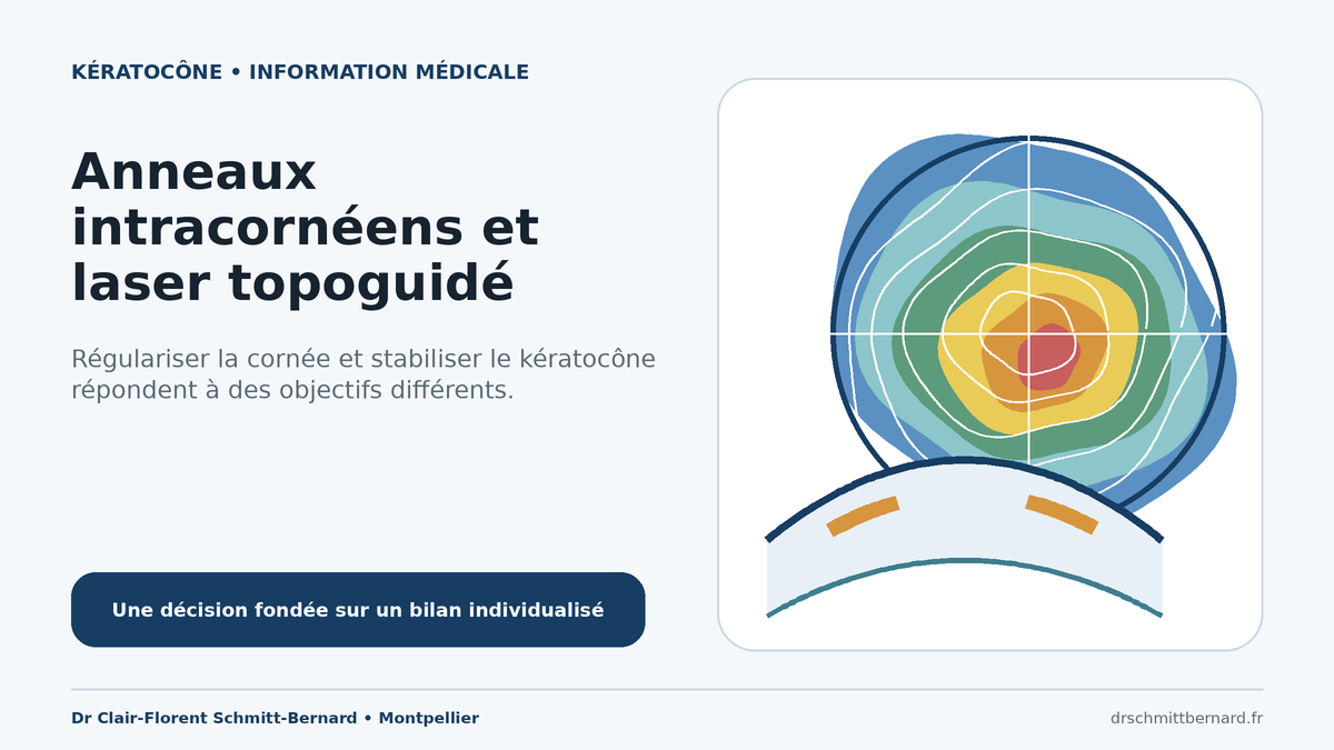
In keratoconus, improving visual quality and stabilising the disease are separate questions. Intracorneal ring segments mechanically alter corneal shape. Topography-guided excimer treatment removes a small amount of tissue according to a personalised map to reduce selected irregularities. When progression is documented, cross-linking has a biomechanical stabilisation objective. Recent evidence supports selecting each procedure according to corneal geometry, thickness and visual function, without promising complete correction. [1,2]
Understanding the three objectives
Keratoconus progressively changes corneal shape and causes irregular astigmatism, sometimes with coma and ghost images. Management may pursue three different objectives: correcting vision with spectacles or contact lenses, slowing progression with corneal cross-linking, or regularising corneal geometry and optics. Intracorneal ring segments and topography-guided PRK mainly address the third objective. They are neither automatic nor interchangeable, and they do not replace ectasia monitoring. [1,2]
Mechanical regularisation with intracorneal ring segments
Intracorneal ring segments are polymer implants, most commonly made of polymethyl methacrylate, placed within the peripheral stroma. Their effect is mechanical: they alter curvature, partially flatten the central area and may partly recenter a decentered cone. No corneal tissue is removed. The number of segments, arc length, thickness and position are selected from topography, tomography, cone location and a nomogram. The intended result is partial regularisation, not restoration of a normal cornea. [2,3]
Implantation is mainly considered when spectacles do not adequately correct irregular astigmatism and rigid contact lenses are intolerable, unstable or insufficient. A clear central cornea and adequate thickness along the planned tunnel are required. A 2024 analysis stresses that eyes with good preoperative corrected acuity have limited potential to gain and may lose lines of vision. The benefit-risk assessment must therefore begin with the patient’s actual functional limitation. [3]
Role of the femtosecond laser
The stromal tunnel can be created mechanically or with a femtosecond laser. An anterior-segment OCT study found more predictable depth with the femtosecond approach. In a comparative cohort, mechanical and refractive complications were less frequent with femtosecond-assisted surgery than with manual dissection, with a marked experience effect in the manual group. These findings support tunnel precision, but they do not remove risk or make the optical response fully predictable. [4,5]
Optical regularisation with topography-guided laser treatment
Topography-guided PRK uses a corneal surface map to programme a personalised ablation. In keratoconus, the aim is not to treat myopia as it would be treated in a normal cornea. The profile is designed primarily to reduce selected irregularities, including coma, while strictly limiting tissue removal. Suitability depends on pachymetry, anterior and posterior tomography, cone location, stability and biomechanical risk. [1,6,7]
When progression is documented, cross-linking retains a separate purpose: stabilising the ectasia. It may be combined with topography-guided treatment in selected protocols, either simultaneously or sequentially. Current evidence does not establish one universal sequence. Corneal condition, predicted residual thickness and the visual objective determine the strategy. [1,2,6]
Available comparative evidence
Published outcomes are encouraging but heterogeneous. A prospective cohort of 45 eyes followed for a mean of 44 months reported better corrected acuity and a greater reduction in higher-order aberrations after topography-guided PRK with accelerated cross-linking than after cross-linking alone. The study was not randomised and the groups were small. [6]
A prospective series of 600 eyes described regularisation and stability at ten years after limited topography-guided ablation with cross-linking, but it had no randomised control group. Another prospective nonrandomised study of 57 eyes compared cross-linking plus ring segments with cross-linking plus topography-guided PRK over four to five years. Uncorrected acuity improved more in the ring group after adjustment, whereas the improvement in corrected acuity did not differ significantly. Baseline characteristics were not identical. These studies therefore do not establish general superiority of either technique. [7,8]
Individualised choice and follow-up
Assessment includes refraction, corrected acuity, slit-lamp examination, topography and tomography with elevation and thickness maps, pachymetry along the planned tunnel, aberrometry and comparison of serial examinations. Central clarity, pupil diameter, contact-lens experience and visual expectations complete the evaluation. The 2026 international consensus supports management organised around the clinical objective and individual risk profile. [1,2]
After ring implantation, halos, glare, migration, extrusion, deposits, infection or insufficient effect may occur. Segments can be removed or exchanged, but explantation does not guarantee exact return to the initial geometry. After topography-guided PRK, epithelial healing, haze, infection, residual correction and tissue removal must be discussed. Spectacles or contact lenses may still be required after either procedure, and long-term monitoring remains necessary. [2,3]
In practice
The decision is not a simple contest between ring segments and laser treatment. The first step is to identify the main problem: ectasia progression, poor optical quality, contact-lens intolerance or a combination of these factors. Management may remain optical, include stabilisation, or add mechanical or topography-guided regularisation. Reproducible measurements and clear information about each option’s limits are essential.
Sources
[1] Global consensus on keratoconus — Cornea, 2026 [2] Evidence-based clinical practice guideline — Clinical Ophthalmology, 2026 [3] Selection criteria for intracorneal ring segments — Eye and Vision, 2024 [4] Predictability of stromal tunnel depth — Journal of Refractive Surgery, 2018 [5] Complications after manual or femtosecond-assisted tunnel creation — Current Eye Research, 2019 [6] Topography-guided PRK with accelerated cross-linking — Frontiers in Medicine, 2024 [7] Ten-year outcomes of topography-guided ablation with cross-linking — Journal of Refractive Surgery, 2023 [8] Ring segments or topography-guided PRK combined with cross-linking — JCRS, 2024 [9] Intracorneal ring segments in paediatric keratoconus: up to ten years — European Journal of Ophthalmology, 2026Frequently asked questions
Do ring segments stop keratoconus?
No. Their main purpose is corneal shape regularisation. Documented progression prompts separate consideration of cross-linking, regardless of the visual effect of the segments. [1,2]
Can topography-guided laser treatment be performed alone?
This depends on stability, thickness and the objective. In progressive ectasia, stabilisation must be addressed separately. Ablation remains limited and is reserved for selected corneas. [1,6]
Are the segments reversible?
They can be removed or exchanged. However, this additional procedure does not guarantee exact restoration of the initial corneal geometry. [2,3]
Which technique gives the best result?
Neither technique is superior for every keratoconus pattern. Cone location, thickness, progression, corrected acuity and contact-lens tolerance guide the choice. [1,8]