Ophthalmology and panorama of Montpellier

Dr Clair-Florent Schmitt-Bernard — Ophthalmic surgeon

  • Corneal diseases and surgery
  • Keratoconus
  • Refractive surgery
  • Therapeutic lasers
Former University Clinical FellowPhD in Science

Choose the appropriate location

Four main areas of expertise

This information presents the main areas of practice. A treatment indication cannot be established without a consultation and an ophthalmic examination.

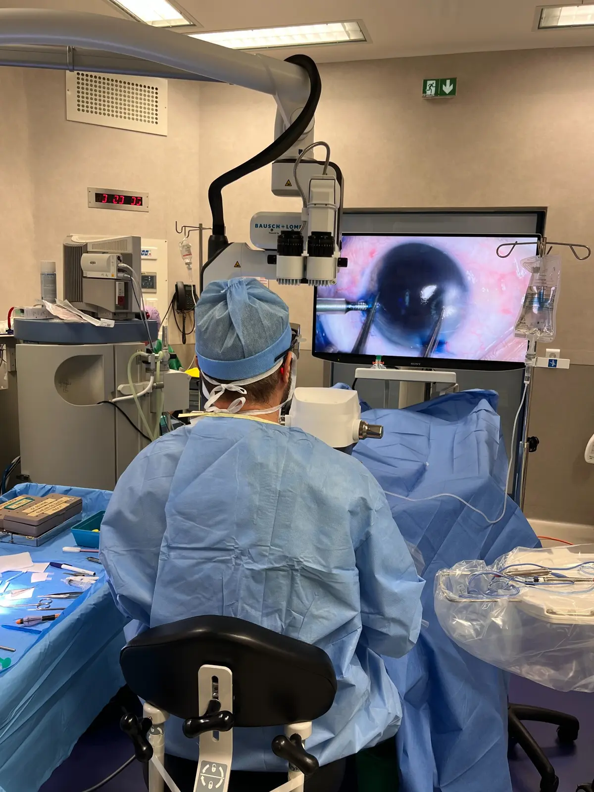
Corneal surgery in an ophthalmic operating theatre

01Cornea

Corneal diseases and surgery

The cornea is the transparent front part of the eye. Changes in its transparency, curvature or structure can reduce vision and may require medical, laser or surgical treatment.

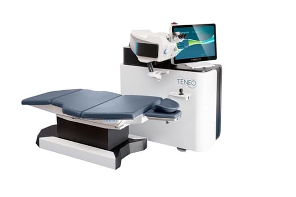
Femtosecond laser cutting. The choice of technique depends on the affected corneal layer, endothelial status, corneal geometry and functional goals. When appropriate, femtosecond laser cutting allows the depth and geometry of tissue planes to be programmed precisely. It does not replace every manual technique, and its value is assessed case by case.

Full-thickness corneal graft

Penetrating keratoplasty replaces the full thickness of the central cornea with a sutured donor graft. It remains indicated when several corneal layers are affected. Femtosecond cutting can create programmed interlocking profiles; however, studies have not demonstrated systematic superiority for every outcome, particularly astigmatism.

Endothelial grafts · UT-DSAEK and DMEK

These procedures selectively replace the posterior cornea when the endothelium fails. UT-DSAEK includes the endothelium, Descemet membrane and an ultra-thin stromal layer. DMEK transplants Descemet membrane and the endothelium.

Anterior lamellar graft · ALTK

ALTK replaces an anterior corneal layer while preserving healthy posterior layers. It may be performed manually or with femtosecond laser assistance. In selected superficial indications, accurate tissue planes may allow a sutureless procedure.

Deep anterior lamellar graft · DALK / DLK

Deep anterior lamellar keratoplasty replaces diseased stroma while preserving Descemet membrane and the endothelium when they remain healthy. It is used particularly for keratoconus and selected stromal disorders.

Bowman layer transplantation

In selected advanced and progressive keratoconus, implantation of a donor Bowman layer within the stroma may reduce or stabilise corneal steepening and defer a more invasive graft. This remains a highly specialised indication.

Amniotic membrane graft

Cryopreserved or lyophilised amniotic membrane may provide biological support for healing in selected tissue defects, ulcerations or conjunctival-corneal reconstructions. The form used depends on the clinical situation.

Pterygium and ocular-surface reconstruction

A pterygium is a fibrovascular growth of conjunctiva extending onto the cornea. Surgery may combine excision with a conjunctival-limbal autograft. Lyophilised amniotic membrane may supplement reconstruction in selected extensive defects or recurrent cases.

Read the full information page →
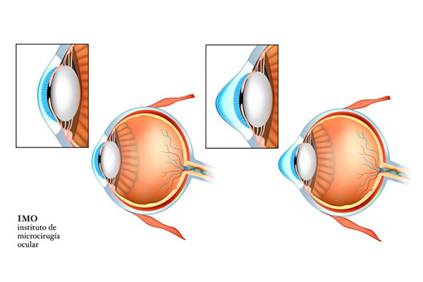
Diagram of corneal deformation caused by keratoconus

02Keratoconus

Keratoconus

Keratoconus is a corneal ectasia in which the cornea progressively thins and deforms, usually asymmetrically. Estimated prevalence varies between populations and according to screening methods.

Mechanism and visual effects

Changes in stromal organisation and resistance lead to corneal protrusion. Astigmatism becomes irregular and is not always fully corrected with spectacles. Screening and comparison of successive examinations are used to identify progression.

Intracorneal rings and topography-guided laser

Segments implanted within the stroma may regularise corneal shape in selected cases. Topography-guided PRK primarily aims to reduce optical irregularities; tissue removal must remain conservative and the procedure may be combined with cross-linking.

Cross-linking and iontophoresis

Cross-linking combines riboflavin and UVA to increase corneal biomechanical resistance and slow documented progression. Iontophoresis uses a low electrical current to promote riboflavin passage through an intact epithelium. By preserving the epithelium, it avoids risks related to epithelial removal, reduces postoperative pain and allows faster corneal recovery.

Hybrid and scleral lenses

Hybrid lenses combine a rigid central zone with a soft skirt. Scleral lenses rest on the sclera and create a fluid reservoir in front of the cornea. They may improve visual quality when spectacles or conventional contact lenses are insufficient.

Phakic ICLs in keratoconus

In selected patients with stable keratoconus, a phakic lens may correct residual myopia or astigmatism without removing corneal tissue. It does not regularise the cornea or treat the ectasia. Stability and ocular anatomy must be assessed.

Read the full information page →
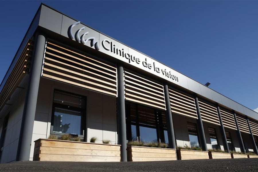
Clinique de la Vision Montpellier

03Refractive surgery

Refractive surgery

25 yearsof refractive-surgery practice

Refractive surgery includes corneal and intraocular procedures designed to change the eye’s optical correction. The choice between PRK, LASIK, SMILE or an ICL can only be made after a complete assessment.

PRK / TRANS-PRK · photorefractive keratectomy

PRK reshapes the corneal surface with an excimer laser after removal of the epithelium. It does not create a flap. Initial recovery is more gradual than with LASIK, and suitability depends on the correction and corneal characteristics.

LASIK

A corneal flap is prepared with a femtosecond laser, after which the stroma is reshaped with an excimer laser. Visual recovery is generally rapid. Corneal thickness, shape and biomechanical stability determine eligibility.

SMILE

A femtosecond laser creates a lenticule within the cornea, which is removed through a small incision without a LASIK flap. The technique is intended for selected myopic and astigmatic corrections.

Procedures combined with cross-linking

In selected fragile corneas or those with borderline parameters, cross-linking may be combined with a refractive procedure. This is not routine: the indication is based on the complete assessment, and available evidence does not support systematic use.

STAAR ICLs

An ICL is a flexible phakic lens placed behind the iris and in front of the natural lens, without removing corneal tissue. It may correct selected myopia and astigmatism when ocular anatomy allows. The assessment includes anterior-chamber and endothelial measurements.

Read the full information page →
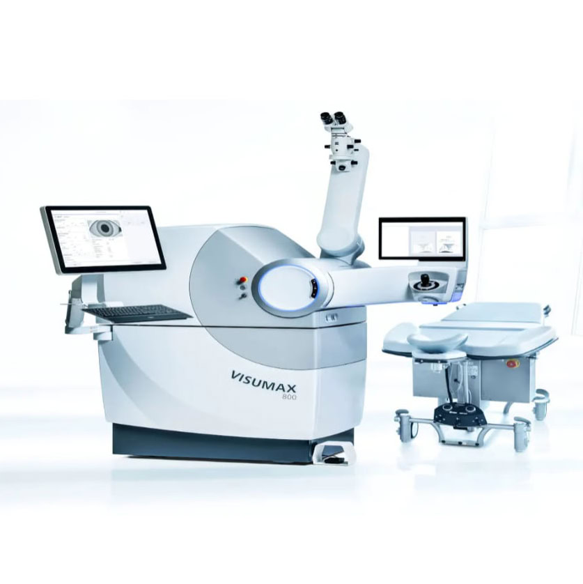
VisuMax 800 femtosecond laser

04Therapeutic corneal lasers

Therapeutic corneal lasers

The excimer laser can remove a programmed amount of corneal tissue. The femtosecond laser creates precise intrastromal tissue planes. Their therapeutic use depends on the disorder and the characteristics of the cornea.

PTK · phototherapeutic keratectomy

PTK uses the excimer laser to remove a controlled superficial depth of cornea. It may be considered for selected superficial corneal dystrophies, superficial opacities or recurrent erosions, with the aim of improving surface regularity or reducing painful recurrences.

Topography-guided PRK

A personalised ablation based on topography may reduce selected corneal irregularities, including in selected keratoconus or after surgery. The indication considers pachymetry, tomography and biomechanical risk; cross-linking may be combined when appropriate.

Femtosecond arcuate incisions

Marked astigmatism may persist after corneal transplantation. Femtosecond-programmed arcuate incisions can selectively relax the steepest meridians. The response varies with healing and residual correction may remain.

Cross-linking uses UVA with riboflavin; it is not a laser treatment.

Read the full information page →

Dr Clair-Florent Schmitt-Bernard

Education
  • Doctor of Medicine · Montpellier I, 1996
  • Specialist qualification in ophthalmology · 1996
  • PhD in Science · Montpellier I, 2001
Cornea
  • Corneal-transplant referral physician at Montpellier University Hospital · 1997–2002
  • Research on TGFBI-associated corneal dystrophies
Practice
  • Private practice in Montpellier since 2002
  • Co-founder of Clinique de la Vision Montpellier · 2012
  • RPPS 10003229605

Facilities and collaborations

Direct access to practice locations, professional collaborations and organisations involved in tissue procurement, preservation and transplantation.

Consultations and procedures in Montpellier

Consultation office

Odysseum Ophtalmologie · Le Farenheit120 avenue Nina Simone · 34000 Montpellier · France+33 4 67 06 06 67