Keratoconus is a corneal ectasia in which the cornea progressively thins and deforms, usually asymmetrically. Estimated prevalence varies between populations and according to screening methods.
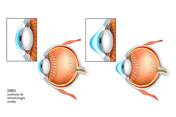
Diagram of corneal deformation caused by keratoconus
Summary
Corneal deformation causes irregular astigmatism, ghost images, halos and reduced visual quality. Topography, tomography, pachymetry and longitudinal assessment help distinguish between visual correction, biomechanical stabilisation and optical regularisation.
Medical information
Principles and techniques
01
Mechanism and visual effects
Changes in stromal organisation and resistance lead to corneal protrusion. Astigmatism becomes irregular and is not always fully corrected with spectacles. Screening and comparison of successive examinations are used to identify progression.
02
Intracorneal rings and topography-guided laser
Segments implanted within the stroma may regularise corneal shape in selected cases. Topography-guided PRK primarily aims to reduce optical irregularities; tissue removal must remain conservative and the procedure may be combined with cross-linking.
03
Cross-linking and iontophoresis
Cross-linking combines riboflavin and UVA to increase corneal biomechanical resistance and slow documented progression. Iontophoresis uses a low electrical current to promote riboflavin passage through an intact epithelium. By preserving the epithelium, it avoids risks related to epithelial removal, reduces postoperative pain and allows faster corneal recovery.
04
Hybrid and scleral lenses
Hybrid lenses combine a rigid central zone with a soft skirt. Scleral lenses rest on the sclera and create a fluid reservoir in front of the cornea. They may improve visual quality when spectacles or conventional contact lenses are insufficient.
05
Phakic ICLs in keratoconus
In selected patients with stable keratoconus, a phakic lens may correct residual myopia or astigmatism without removing corneal tissue. It does not regularise the cornea or treat the ectasia. Stability and ocular anatomy must be assessed.
Progression is assessed by comparing successive refraction, topography, tomography and corneal-pachymetry measurements. Documented changes in corneal shape or thickness may lead to consideration of stabilising treatment.
Does cross-linking improve vision?
The main aim of cross-linking is to slow documented progression by increasing corneal biomechanical resistance. It does not systematically correct irregular astigmatism; visual correction may still require contact lenses or a selected complementary procedure.
Which contact lenses may help vision in keratoconus?
Depending on corneal geometry, rigid, hybrid or scleral lenses may be considered. Scleral lenses create a fluid reservoir in front of the cornea and may improve visual quality when spectacles are insufficient.