Keratoconus is a corneal ectasia: the cornea progressively thins and changes shape, often asymmetrically. This can cause irregular astigmatism, ghost images, halos, and reduced visual quality. When progression is objectively documented, corneal cross-linking may be considered to increase stromal biomechanical resistance and slow further change. Its primary purpose is disease stabilization; it does not replace glasses or contact lenses and does not reliably correct vision. [1–4]
Why must progression be documented before treatment?
A cornea with keratoconus does not necessarily progress at the same rate throughout life. Decisions therefore rely on comparable serial examinations: refraction and corrected visual acuity, corneal topography or tomography, keratometry, and pachymetry. A consistent increase in steepness, posterior shape change, or thinning may help demonstrate progression. There is no single universal threshold, however: measurement quality and repeatability, age, and the clinical context must be assessed together. The 2026 international consensus emphasizes individualized management. [1]
This distinction matters: cross-linking is a stabilizing treatment for progressive ectasia, not an automatic preventive procedure for every suspicious topography. In younger patients, in whom progression may be faster, monitoring is generally closer and treatment may be discussed earlier when supported by objective findings. [1,3]
How does corneal cross-linking work?
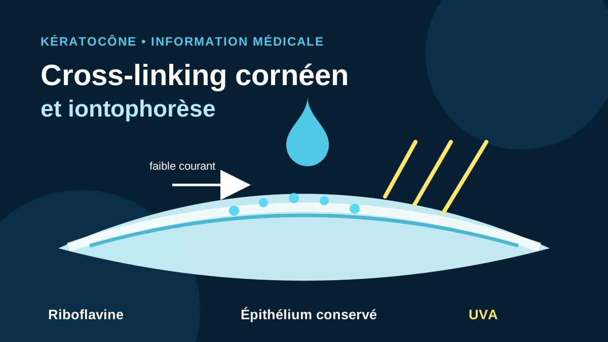
Treatment combines a riboflavin solution, or vitamin B2, with controlled ultraviolet A irradiation. Photoactivation of riboflavin initiates reactions that increase bonding within the stromal matrix, making the treated cornea mechanically more resistant. Cross-linking is therefore not refractive laser surgery: it does not intentionally reshape the cornea to remove an optical prescription. [2,8]
In the historical epithelium-off protocol, irradiance is 3 mW/cm² for 30 minutes. Total dose is calculated as follows: 3 mW/cm² × 1,800 seconds = 5,400 mJ/cm², or 5.4 J/cm². Accelerated protocols use higher irradiance for a shorter period. An equivalent energy dose does not necessarily mean complete biological equivalence, because oxygen, riboflavin diffusion, and irradiation mode also matter. [8]
Epithelium-off cross-linking: the longest evidence base
The “epi-off” protocol temporarily removes the central epithelium to facilitate stromal riboflavin penetration. Randomized trials have shown a lower risk of progression than observation in patients with progressive keratoconus. Cohorts followed for up to ten years document sustained stabilization in most patients, while also showing that renewed progression can occur and that follow-up remains necessary. [2–4]
Epithelial removal accounts for part of the recovery: pain, tearing, light sensitivity, and blurred vision while healing occurs. A bandage contact lens and topical medication are commonly prescribed. Reported complications include delayed epithelial healing, corneal haze, infection and, less commonly, persistent loss of corrected visual acuity. Corneal thickness and ocular surface status must be assessed before selecting a protocol. [3,8]
What does iontophoresis change?
Iontophoresis uses a low-intensity electrical current to promote the passage of ionized riboflavin through an intact epithelium. This “epi-on” approach avoids a central epithelial wound. Studies generally report less postoperative pain and faster early recovery. Its potential benefit therefore concerns treatment tolerance, without removing the need for careful patient selection or tomographic follow-up. [5–7]
The trade-off relates to the epithelial barrier, which may limit stromal penetration of riboflavin, oxygen, and UVA. In a two-year randomized trial, both groups had favorable clinical outcomes, but corneal apex flattening was greater after standard cross-linking. A five-year iontophoresis cohort reported ABCD-defined re-progression in 25.9% of eyes; this figure comes from one study and should not be extrapolated to every protocol or patient. [5,6]
The meta-analysis published in February 2026 included 14 studies and 629 patients. It suggests that both approaches can stabilize disease, with greater corneal flattening after epi-off treatment and a generally more favorable postoperative experience after iontophoresis. Heterogeneity in riboflavin formulations, irradiation parameters, patient age, and progression criteria requires cautious interpretation. [7]
How is a protocol selected?
The choice is not between a “modern” and an “old” technique. It balances the strength of evidence, expected biomechanical effect, corneal thickness and geometry, age, rate of progression, healing capacity, and follow-up constraints. Epi-off treatment remains the reference comparator and has the strongest long-term evidence. Iontophoresis may be considered in selected situations after discussing its tolerance advantages and the remaining uncertainty about comparative long-term effectiveness. [1,4,7]
After treatment: monitoring and visual correction
Stability cannot be judged from a single symptom or examination. Follow-up combines corneal examination with the same reproducible measurements used before treatment. Vision may fluctuate for several weeks or months. Even when keratoconus is stable, glasses or rigid, hybrid, or scleral lenses may remain necessary. In selected cases, other procedures may aim to improve optical regularity, but that objective differs from cross-linking. [1,4]
In summary, cross-linking addresses the risk of progression, while optical correction addresses visual quality. Protocol selection must remain individualized and should include a clear discussion of expected benefits, limitations, and risks.
Sources
[1] International keratoconus consensus — Cornea, 2026 [2] Three-year randomized trial — Ophthalmology, 2014 [3] KERALINK trial — Health Technology Assessment, 2021 [4] Ten-year epithelium-off outcomes — Cornea, 2024 [5] Two-year randomized trial of iontophoresis versus standard cross-linking — JCRS, 2019 [6] Five-year iontophoresis outcomes — International Ophthalmology, 2023 [7] Epithelium-off versus iontophoresis meta-analysis — International Ophthalmology, 2026 [8] Documented standard-protocol parameters — FDAFrequently asked questions
Does cross-linking make keratoconus disappear?
No. It aims to slow documented progression. The cornea remains keratoconic, and optical correction is often still required.
Is iontophoresis as effective as epi-off treatment?
Evidence indicates better early tolerance, but the protocols are not interchangeable in every situation. Epi-off cross-linking retains the strongest long-term evidence base.
How is treatment success assessed?
By comparing serial examinations: topography or tomography, pachymetry, refraction, and corrected visual acuity. Long-term follow-up is required.
Is cross-linking a laser procedure?
No. It uses riboflavin activated by UVA. Its biomechanical goal differs from excimer-laser refractive photoablation.