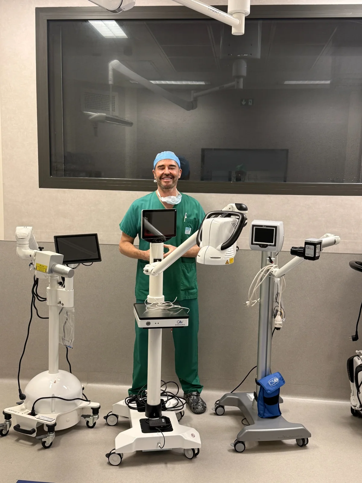
Three generations of devices
A recently published Instagram post looks back on fifteen years of corneal cross-linking for progressive keratoconus. Three generations of devices represent the transition from early protocols to optimised procedures and then to theranostic monitoring. This timeline is a professional account, not a clinical comparison of the devices.
What theranostic monitoring measures
Cross-linking combines riboflavin with controlled ultraviolet A irradiation to increase corneal biomechanical resistance. Its purpose is to slow objectively documented keratoconus progression. Theranostic monitoring adds optical measurements during treatment: the studied device estimates corneal riboflavin concentration, monitors its UVA-mediated photodegradation and calculates a score intended to estimate treatment effect during the procedure. [1–3]
Exclusive use of iontophoresis in my practice
In my practice, cross-linking is performed exclusively with iontophoresis and without epithelial removal. A low-intensity electrical current delivers riboflavin through an intact epithelium. Avoiding a central epithelial wound markedly reduces risks specifically related to de-epithelialisation — substantial pain, delayed healing, surface infection and haze — and usually allows a faster return towards the pre-procedure level of vision. It does not remove every risk of cross-linking or the need for long-term tomographic follow-up. [5–7]
What the first clinical data show
A multicentre randomised clinical trial in 50 patients assessed whether the combined riboflavin and theranostic scores could predict Kmax flattening at one year. The trial provides favourable clinical evidence, but it remains limited in size and concerns a defined device, protocols and thresholds. It should not be interpreted as a guarantee of individual stabilisation. [3]
A useful measurement with limitations
The measurement is based on riboflavin fluorescence. A 2026 critical paper notes that this optical signal may also be influenced by light scattering, absorption, hydration and tissue changes during irradiation. The score is therefore a device-specific intraoperative indicator; serial corneal topography or tomography remains necessary to assess actual keratoconus progression. [4]
Fifteen years of collaboration
The post also refers to long-term collaboration with Horus Pharma and Clinique Saint-Roch in Montpellier. This professional history is presented as one of progress, measurement and procedural personalisation, without changing the indications for cross-linking or the requirements for patient selection and follow-up.
Sources
Instagram post behind this news item [1] Non-invasive riboflavin measurement and real-time estimation — Journal of Biophotonics, 2018 [2] Preclinical evidence for theranostic-guided cross-linking — Journal of Biophotonics, 2022 [3] Multicentre randomised clinical trial — Ophthalmology Science, 2024 [4] Critical discussion of fluorescence-based quantification — Journal of Biophotonics, 2026 [5] Randomised trial of iontophoresis versus standard cross-linking — JCRS, 2019 [6] Five-year iontophoresis outcomes — International Ophthalmology, 2023 [7] Meta-analysis of iontophoresis versus epi-off cross-linking — International Ophthalmology, 2026Frequently asked questions
Does the theranostic score prove that keratoconus has been stabilised?
No. It estimates during the procedure the effect associated with riboflavin and its UVA activation. Stabilisation is subsequently assessed by comparing reproducible corneal examinations over time. [3,4]
Does theranostic monitoring change the indication for cross-linking?
No. Cross-linking is still considered for objectively documented progressive keratoconus after assessment of the cornea and its thickness.